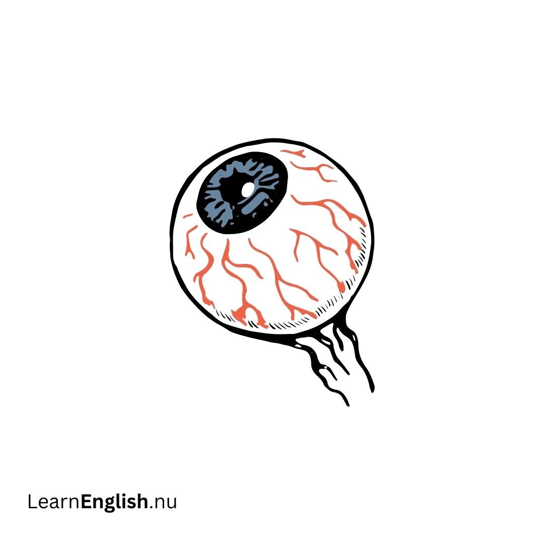
Anatomy of the Eye in Swedish
Understanding the anatomy of the eye is essential for grasping how we see the world. This guide covers the basic structures and their functions, helping you learn the main components of the human eye and their roles in vision.
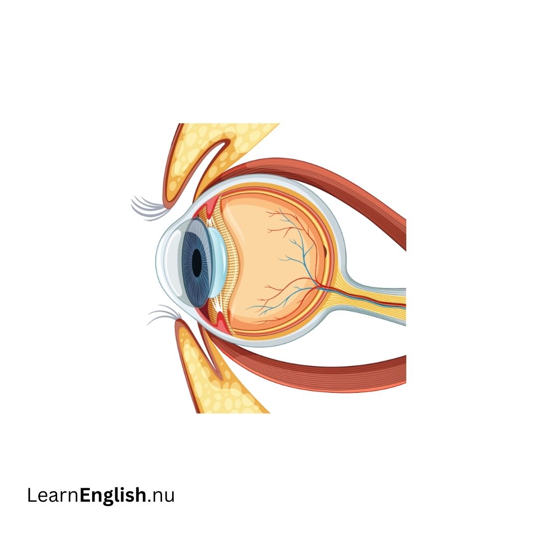
Key Structures of the Eye
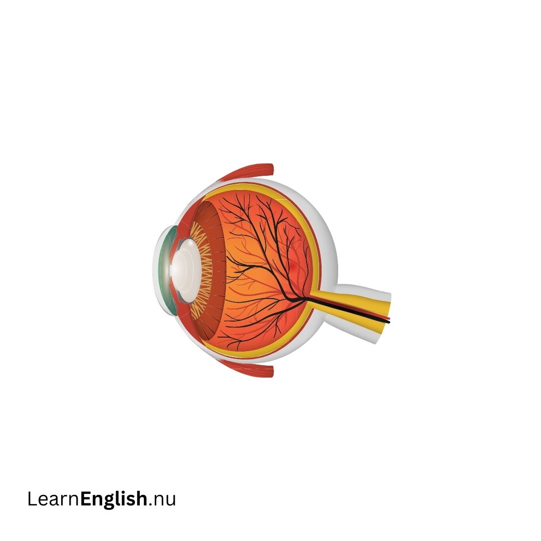
- Choroid The choroid is a layer of blood vessels located between the retina and the sclera at the back of the eye. It supplies nutrients to the retina and helps maintain its health.
- Ciliary Body Situated behind the iris, the ciliary body contains muscles that adjust the lens's focus, allowing us to see objects clearly at various distances.
- Cornea The cornea is the eye's clear, dome-shaped front window. It focuses light onto the retina and can be reshaped with laser surgery to correct vision.
- Fovea The fovea is the central part of the macula and is responsible for sharp, detailed central vision.
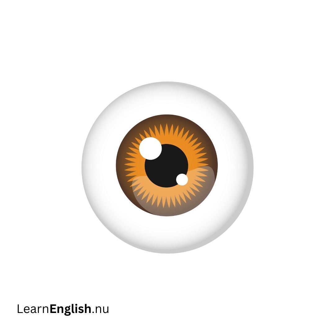
- Iris The iris is the colored part of the eye. It regulates the amount of light entering the eye by adjusting the size of the pupil based on lighting conditions.
- Lens The transparent lens focuses light onto the retina. It can be replaced if needed, such as during cataract surgery, and its ability to focus decreases with age.
- Macula The macula, located in the retina, contains light-sensitive cells that allow for clear, detailed central vision. Age-related macular degeneration (ARMD) affects this area as we age.
- Optic Nerve The optic nerve is a bundle of over a million nerve fibers that transmits visual information from the retina to the brain, where images are processed and interpreted.
- Pupil The pupil is the dark opening in the center of the iris. It changes size to control the amount of light entering the eye, similar to the aperture in a camera.
- Retina The retina is the light-sensitive nerve layer lining the back of the eye. It converts light into electrical impulses sent to the brain through the optic nerve.
- Sclera The sclera is the white, outer coating of the eye that provides structure and protection.
- Vitreous Humor The vitreous humor is a clear, gelatinous substance filling the eye's central cavity, maintaining its shape and optical properties.
| Structure |
Function |
Swedish Translation |
Swedish Function |
| Choroid |
Supplies nutrients to the retina and supports its health. |
Choroid |
Försörjer näthinnan med näring och stödjer dess hälsa. |
| Ciliary Body |
Adjusts the lens focus for clear vision. |
Ciliarkropp |
Justerar linsens fokus för tydlig syn. |
| Cornea |
Focuses light onto the retina and can be reshaped by surgery. |
Hornhinna |
Fokuserar ljus på näthinnan och kan omformas vid operation. |
| Fovea |
Provides sharp, detailed vision at the center of the visual field. |
Fovea |
Ger skarp, detaljerad syn i mitten av det synliga området. |
| Iris |
Regulates light entry by adjusting the pupil size. |
Iris |
Reglerar ljusinsläppet genom att justera pupillens storlek. |
| Lens |
Focuses light on the retina and can be replaced if needed. |
Lins |
Fokuserar ljus på näthinnan och kan bytas ut vid behov. |
| Macula |
Allows for detailed central vision and can deteriorate with age. |
Makula |
Möjliggör detaljerad central syn och kan försämras med åldern. |
| Optic Nerve |
Transmits visual information from the retina to the brain. |
Synnerv |
Överför visuell information från näthinnan till hjärnan. |
| Pupil |
Controls light entry by changing size. |
Pupill |
Kontrollerar ljusinsläpp genom att ändra storlek. |
| Retina |
Converts light into electrical impulses for the brain. |
Näthinna |
Omvandlar ljus till elektriska impulser för hjärnan. |
| Sclera |
Provides structure and protection for the eye. |
Sclera |
Ger struktur och skydd åt ögat. |
| Vitreous Humor |
Maintains eye shape and optical properties. |
Glaskropp |
Bibehåller ögats form och optiska egenskaper. |
How the Eye Works
Understanding how our eyes work helps us appreciate the complexity of vision. This guide explains the basic process of sight and how our eyes and brain work together to help us see.
How Vision Works
Our sense of sight is a crucial part of our sensory experience, working closely with other senses and parts of our anatomy. The process of seeing involves light traveling through the eye and being interpreted by the brain.
The Process of Seeing
- Light Entry and Focus (Ljusinsläpp och Fokus) Light enters the eye through the cornea, the clear front surface. It then passes through the lens, which focuses the light onto the retina at the back of the eye. This process is similar to how a camera lens focuses light onto film or a sensor. (Ljus passerar genom hornhinnan, den klara ytan på framsidan av ögat. Det går sedan genom linsen, som fokuserar ljuset på näthinnan längst bak i ögat. Denna process är liknande hur en kamerlins fokuserar ljus på film eller en sensor).
- Image Conversion ( Bildomvandling) The retina contains special cells that absorb the light and convert it into electrochemical signals. These signals travel along the optic nerve to the brain, where they are processed and interpreted as images. (Näthinnan innehåller speciella celler som absorberar ljuset och omvandlar det till elektrokemiska signaler. Dessa signaler färdas längs med synnerven till hjärnan, där de bearbetas och tolkas som bilder).
- Adjusting Light Intake (Justering av Ljusinsläpp) Just as a camera shutter controls the amount of light hitting the film, the iris and pupil in the eye regulate light entry. In low light, the pupils dilate to let in more light, while in bright conditions, they constrict to reduce light intake. (Precis som en kamerastängning kontrollerar mängden ljus som träffar filmen, reglerar iris och pupill i ögat ljusinsläppet. Vid svagt ljus vidgas pupillerna för att släppa in mer ljus, medan de vid starkt ljus dras ihop för att minska ljusinsläppet).
- Focusing Vision (Fokusering av Synen)The eye's lens focuses on objects at varying distances. If vision needs correction, glasses, contact lenses, or artificial lenses help by adjusting the focus to ensure clear vision. (Ögats lins fokuserar på objekt på olika avstånd. Om synen behöver korrigeras hjälper glasögon, kontaktlinser eller konstgjorda linser genom att justera fokus för att säkerställa tydlig syn).
| Step |
Description |
Swedish Translation |
| Light Entry and Focus |
Light passes through the cornea and lens, focusing on the retina. |
Ljusinsläpp och Fokus Ljus passerar genom hornhinnan och linsen, och fokuserar på näthinnan. |
| Image Conversion |
Retina converts light into signals sent to the brain via the optic nerve. |
Bildomvandling Näthinnan omvandlar ljus till signaler som skickas till hjärnan via synnerven. |
| Adjusting Light Intake |
Iris and pupil adjust to control light entry, similar to a camera shutter. |
Justering av Ljusinsläpp Iris och pupill justerar ljusinsläppet, liknande en kamerastängning. |
| Focusing Vision |
Lens focuses light, with corrective lenses used for clear vision. |
Fokusering av Synen Linsen fokuserar ljuset, och korrigerande linser används för tydlig syn. |
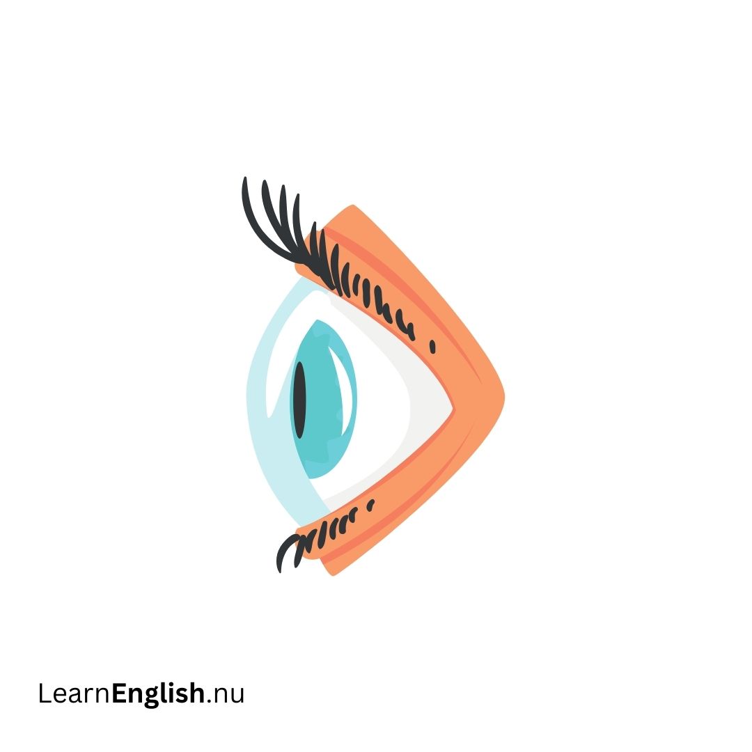
Understanding the Surface of the Eye in Swedish
The surface of the eye plays a crucial role in maintaining clear and comfortable vision. This guide will walk you through the main components of the eye's surface, how tears work, and the functions of the front and back parts of the eye.
The Surface of the Eye
The surface of the eye, including the inner eyelids, is covered by a clear membrane known as the conjunctiva (konjunktiva). This membrane plays a key role in protecting and lubricating the eye.
The Conjunctiva and Tear Film
Conjunctiva (Konjunktiva) The conjunctiva is a thin, transparent membrane that covers the white part of the eye (sclera) and the inner surface of the eyelids. It helps keep the eye moist and protected.
Tear Film Layers (Tårfilmens Lager) Tears are essential for keeping the eye lubricated and comfortable. The tear film consists of three layers:
- Mucous Layer (Slemhinna): Produced by the conjunctiva, this layer helps spread the tears evenly over the eye.
- Watery Layer (Vattenlager): Secreted by the lacrimal gland located above the outer edge of the eyebrow, this layer provides the main moisture.
- Oil Layer (Oljelager): Created by the meibomian glands, this layer prevents tears from evaporating too quickly.
The Front of the Eye
The front part of the eye is essential for focusing light. It includes the cornea (hornhinna), anterior chamber (främre kammare), iris (iris), pupil (pupill), and lens (lins).
Components and Functions
- Cornea and Anterior Chamber (Hornhinna och Främre Kammare) The cornea is the clear, dome-shaped front part of the eye that focuses light. Behind it is the anterior chamber, filled with aqueous humor (kammarvätska). This fluid maintains eye pressure and drains through the drainage angle (dräneringsvinkel).
- Iris and Pupil (Iris och Pupill) The iris is the colored part of the eye that controls the size of the pupil, adjusting light entry. The pupil is the dark center that changes size to regulate how much light reaches the retina.
- Lens (Lins) The lens is located directly behind the pupil and focuses light onto the retina (näthinna). It changes shape to help focus on objects at different distances. The lens capsule (linsens kapsel) holds it in place, and replacement lenses can be inserted into the capsule if needed.
The Back of the Eye
The back of the eye is responsible for processing light and transmitting visual information to the brain. It includes the vitreous cavity (glaskroppens hålighet), retina (näthinna), macula (makula), and photoreceptors (fotoreceptorer).
Components and Functions
- Vitreous Humor (Glaskroppsvätska) The vitreous cavity, filled with vitreous humor, lies between the lens and the retina. This gel-like substance helps maintain the eye's shape.
- Retina and Macula (Näthinna och Makula) Light focused by the cornea and lens passes through the vitreous humor to the retina, which is the light-sensitive tissue lining the back of the eye. The macula, a specialized area of the retina, provides detailed central vision, while the peripheral retina handles side vision.
- Photoreceptors (Fotoreceptorer) The retina contains photoreceptor cells called rods (stavar) and cones (koner). Rods detect black and white and enable night vision, while cones detect color and provide detailed central vision. These cells convert light into electrical impulses sent through the optic nerve (synnerv) to the brain.
| Part of the Eye |
Function |
Swedish Translation |
Swedish Function |
| Conjunctiva |
Covers and protects the eye surface and inner eyelids. |
Konjunktiva |
Täcker och skyddar ögats yta och inre ögonlock. |
| Tear Film Layers |
Keeps the eye lubricated with mucous, watery, and oil layers. |
Tårfilmens Lager |
Håller ögat fuktigt med slemhinna, vatten- och oljelager. |
| Cornea |
Focuses light into the eye. |
Hornhinna |
Fokuserar ljus in i ögat. |
| Anterior Chamber |
Contains aqueous humor and maintains eye pressure. |
Främre Kammare |
Innehåller kammarvätska och upprätthåller ögontrycket. |
| Iris and Pupil |
Controls light entry by adjusting the pupil size. |
Iris och Pupill |
Reglerar ljusinsläpp genom att justera pupillens storlek. |
| Lens |
Focuses light onto the retina and changes shape for focusing. |
Lins |
Fokuserar ljus på näthinnan och ändrar form för att fokusera. |
| Vitreous Humor |
Maintains eye shape and provides support. |
Glaskroppsvätska |
Upprätthåller ögats form och ger stöd. |
| Retina |
Converts light into electrical impulses sent to the brain. |
Näthinna |
Omvandlar ljus till elektriska impulser som skickas till hjärnan. |
| Macula |
Provides detailed central vision. |
Makula |
Ger detaljerad central syn. |
| Photoreceptors |
Rods and cones convert light into energy for vision. |
Fotoreceptorer |
Stav- och konceller omvandlar ljus till energi för syn. |